In my two recent blogs, I wrote about ‘clubfeet’ (talipes) and infant ‘wryneck’ (torticollis). Both are neonatal conditions that are often related to baby being ‘packaged’ in a certain way inside the womb. To complete the series, I am writing about another related ‘packaging’ condition that we as paediatricians have to check in all newborns before they leave the hospital. This condition is called developmental dysplasia of the hip (DDH), or hip dysplasia.
How common is hip dysplasia?
The incidence of DDH is approximately 1 in 100 live births screened clinically, and up to 8 in 100 infants screened by ultrasound (1). In my clinical practice, I see DDH more frequently than I see clubfeet or torticollis. Don’t fret though, it is important to note that DDH comes in a spectrum of severity and that it is treatable. As previously mentioned in my other blogs, infants with clubfeet and torticollis may have DDH as well, so the three conditions would be closely monitored if your baby has one or the other.
Diagnosis of hip dysplasia
Ok, so let’s talk about the hip joint. Make a fist with your right hand and cover your right fist with your left hand. The hip joint is made up of the round head of the top of the femur (the big and long thigh bone); in this case your right fist, being cupped into a socket of the pelvis called the acetabulum; in this case your left covering hand. These ball and socket joints are supported and connected by muscles, tendons and ligaments. Think of them like sticky tape that holds the joint together. Unlike adults, newborn’s bones are soft and pliable (think of bendable pool noodles versus rigid lead pipe).
Developmental dysplasia of the hip occurs when there is abnormal development to this ball and socket joint structure making it unstable. The degree of hip instability varies from one baby to another. You may hear these words being mentioned by your doctor. Subluxation occurs if the ball (femoral head) can be partially displaced out of the cup (acetabulum). Dislocatable hip is when the ball (femoral head) may be displaced from the cup (acetabulum) with manoeuvres. Dislocated hip is when the ball (the femoral head) is completely outside the acetabulum. This hand out from the Royal Children’s Hospital has a really good illustration of hip dysplasia.
Risk factors associated with hip dysplasia
We know that there are some risk factors associated with DDH. This can be divided further into genetic and environmental.
Genetically, study has shown that DDH is twelve times more likely in a baby with family history of DDH (either parents or siblings). During pregnancy, pregnant women release hormones that help relax their ligaments at birth. These hormones are circulated through the baby’s blood as well and is thought to be the reason why girls are four times more likely to have DDH as they are more sensitive to these hormones compared to boys.
Environmentally, think of it as there was not enough space inside the womb, putting more pressure on the hip joint to make it more unstable during its development. DDH is more common in first born babies (‘tighter’ womb and delivery passages), breech baby, baby over 4 kg (bigger size), baby who is overdue by more than two weeks (bigger size), baby who has a low level of amniotic fluid in the womb, and multiple pregnancy (not roomy enough).
Due to the way that babies normally lie inside the womb, DDH commonly affects the left hip more than the right, but it can also affect both. Having a dislocated hip as an adult would be super painful but not in neonates.
Screening for hip dysplasia
As with anything else, early diagnosis and treatment is always better. Your paediatrician will check your baby’s hip regardless of any risk factor before you leave the hospital. If the initial examination is normal but there is a known high risk factor (particularly breech baby and positive family history), then your paediatrician will arrange hip ultrasound at around the six week mark.
If there is abnormal examination with or without risk factors, most paediatricians will arrange hip ultrasound before your baby gets discharged. If the ultrasound result is abnormal then referral to an orthopaedic surgeon would be done before you leave the hospital.
Treatment of hip dysplasia
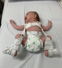
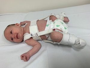
The treatment of DDH varies depending on severity. If it’s really mild, some surgeons might decide to monitor the hip with serial examination and ultrasound, giving time for the ‘sticky tape’ to become stronger. In more severe cases, splinting with braces is the next mode of treatment. The braces would need to be worn for 6-12 weeks with treatment being monitored by the orthopaedic surgeon. The commonest brace used in DDH is the Pavlik Harness.
If splinting does not work or if DDH is diagnosed quite late (at or after three months of age), surgical treatment with subsequent hip plaster cast (spica) might be needed.
Late diagnosis of hip dysplasia
Serial hip examinations by your GP, midwives and paediatrician are required at every well-baby check. There are cases where DDH has been missed. This is usually more frequent in children with no known risk factors. After three months of age, if you feel that your child has a stiff hip joint, uneven skin folds on their groin and thigh, or legs that are different in length, mention this to your health care providers. Long term consequences of undiagnosed DDH leads to pain in the hip, knee and lower back and gait abnormalities (waddling, leg turned outward limping walk) in older child. In the long run, untreated DDH will cause degenerative changes of the hip joint in adulthood.
Swaddling and baby carriers
Babies hip joints are rather malleable for the first couple of months. This is why it is important to adopt a safe swaddling and baby carrying method to prevent putting pressure on your baby’s hip joint. Ensuring that when swaddling baby the hips and knees are not held fully straight with legs brought together for a long time, and when carrying baby in a carrier, to have the hips spread apart naturally to the side. Please take a look at these links for video instruction and diagram of safe swaddling and baby carrying method.

If you feel you require paediatric advice for your child please contact us.
For more qualified, easy-to-understand specialist paediatric information visit Paedicare’s blog.
1. American Academy of Pediatrics. Clinical Practice Guideline: Early detection of developmental dysplasia of the hip. Pediatrics 2000; 105:896-905